File:Trophozoites of Entamoeba histolytica with ingested erythrocytes.JPG
No higher resolution available.
Trophozoites_of_Entamoeba_histolytica_with_ingested_erythrocytes.JPG (282 × 198 pixels, file size: 27 KB, MIME type: image/jpeg)
| This is a file from the Wikimedia Commons. The description on its description page there is shown below.
|
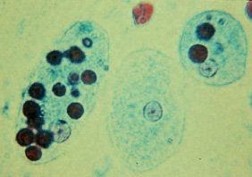
| Description | Trophozoites of Entamoeba histolytica with ingested erythrocytes (trichrome stain). The ingested erythrocytes appear as dark inclusions. Erythrophagocytosis is the only characteristic that can be used to differentiate morphologically E. histolytica from the nonpathogenic E. dispar. In these specimens, the parasite nuclei have the typical small, centrally located karyosome, and thin, uniform peripheral chromatin. | |||
| Source | DPD CDC http://www.dpd.cdc.gov/dpdx/images/ParasiteImages/A-F/Amebiasis/E_histol_trophs_F.JPG | |||
| Author | ||||
| Permission (Reusing this file) |
|
File history
Click on a date/time to view the file as it appeared at that time.
| Date/Time | Dimensions | User | Comment | |
|---|---|---|---|---|
| current | 06:38, 29 April 2006 | 282 × 198 (27 KB) | Patho | {{Information| |Description= Trophozoites of Entamoeba histolytica with ingested erythrocytes (trichrome stain). The ingested erythrocytes appear as dark inclusions. Erythrophagocytosis is the only characteristic that can be used to differentiate morpho |
File usage
The following 4 pages use this file: