File:Animal Cell.svg

Size of this PNG preview of this SVG file: 800 × 462 pixels. Other resolutions: 320 × 185 pixels | 640 × 369 pixels | 1,024 × 591 pixels | 1,280 × 739 pixels | 1,405 × 811 pixels.
Original file (SVG file, nominally 1,405 × 811 pixels, file size: 457 KB)
| This is a file from the Wikimedia Commons. The description on its description page there is shown below.
|
Summary
| Description |
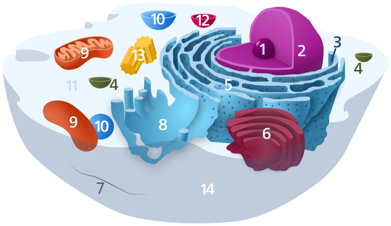
English: A reworked version of File:Biological_cell.svg.
Diagram of a typical animal cell. Organelles are labelled as follows:
العربية: رسم تخطيطي للخلية الحيوانية
Català: Dibuix esquemàtic d'una cèl·lula animal típica:
Español: Diagrama de una célula animal típica:
ਪੰਜਾਬੀ: ਕਿਸੇ ਮਿਸਾਲੀ ਜਾਨਵਰ ਦੇ ਕੋਸ਼ਾਣੂ ਦਾ ਚਿੱਤਰ:
Svenska: Schematisk bild över en typisk eukaryot cell, som visar cellens subcellulära komponenter. Organeller:
Deutsch: Organisation einer typischen eukaryotischen Tierzelle:
|
|||
| Date | ||||
| Source | Own work | |||
| Author | Kelvinsong | |||
| Permission (Reusing this file) |
I, the copyright holder of this work, hereby publish it under the following license:
|
File history
Click on a date/time to view the file as it appeared at that time.
| Date/Time | Dimensions | User | Comment | |
|---|---|---|---|---|
| current | 07:47, 17 November 2022 | 1,405 × 811 (457 KB) | TheBartgry | Reverted to version as of 00:21, 10 December 2012 (UTC) showing continuity between nuclear membrane and ER is useful |
File usage
The following page uses this file: