Atomic force microscope
Atomic force microscopes (AFMs) are a type of microscope. AFMs provide pictures of atoms on or in surfaces. Like the scanning electron microscope (SEM), the purpose of the AFM is to look at objects on the atomic level. In fact, the AFM may be used to look at individual atoms.[1] It is commonly used in nanotechnology.
The AFM can do some things that the SEM cannot do. The AFM can provide higher resolution than the SEM. Further, the AFM does not need to operate in a vacuum. In fact, the AFM can operate in ambient air or water, so it can be used to see surfaces of biological samples like living cells.
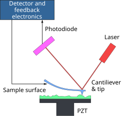
The AFM works by employing an ultra-fine needle attached to a cantilever beam. The tip of the needle runs over the ridges and valleys in the material being imaged, "feeling" the surface. As the tip moves up and down due to the surface, the cantilever deflects. In one basic configuration, a laser shines on the cantilever at an oblique angle, and allows for the direct measurement of the deflection in the cantilever by simply changing the angle of incidence for the laser beam. In this way, an image may be created revealing the configuration of the molecules being imaged by the machine.[2]
There are many different operating modes for an AFM. One is the "contact mode", where the tip is simply moved across the surface and the cantilever deflections are measured. Another mode is called "tapping mode", because the tip is tapped against the surface as it travels along. By controlling how hard the tip is tapped, the AFM can move away from the surface when the needle feels a ridge, so that it will not hit against the surface when it moves across. This mode is also useful for biological samples, because it is less likely to damage a soft surface.[3] These are the basic modes most commonly used. However there are different names and methods such as "intermittent contact mode", "non-contact mode", "dynamic" and "static" modes and more, but these are often variations on the above described tapping and contact modes.
Atomic Force Microscope Media
An AFM generates images by scanning a small cantilever over the surface of a sample. The sharp tip on the end of the cantilever contacts the surface, bending the cantilever and changing the amount of laser light reflected into the photodiode. The height of the cantilever is then adjusted to restore the response signal, resulting in the measured cantilever height tracing the surface.
- Error missing media source
Operating principle of an atomic force microscope
Atomic force microscope topographical scan of a glass surface. The micro and nano-scale features of the glass can be observed, portraying the roughness of the material. The image space is (x,y,z) = (20 μm × 20 μm × 420 nm).
Single polymer chains (0.4 nm thick) recorded in a tapping mode under aqueous media with different pH.
Model for AFM water meniscus
AFM beam-deflection detection
Related pages
References
- ↑ 1. Sugimoto, Y., et al., Chemical identification of individual surface atoms by atomic force microscopy. Nature, 2007. 446(7131): p. 64-67.
- ↑ Binnig, G., C.F. Quate, and C. Gerber, Atomic Force Microscope. Physical Review Letters, 1986. 56(9): p. 930.
- ↑ Weisenhorn, A.L., et al., Deformation and height anomaly of soft surfaces studied with an AFM. Nanotechnology, 1993. 4(2): p. 106-113.


