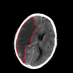
Cerebral infarction
A cerebral infarction is an area of necrotic tissue in the brain caused from a blockage or narrowing in the arteries supplying blood and oxygen to the brain.
The lack of oxygen due to the low blood supply causes an ischemic stroke that can result in an infarction if the blood flow is not restored within a relatively short period of time. The blockage can be due to a thrombus, an embolus or an atheromatous stenosis of one or more arteries.[1]
Which arteries are problematic will determine which areas of the brain are affected (infarcted). These varying infarcts will produce different symptoms and outcomes. About one third will prove fatal.
Cerebral Infarction Media
Hemodynamic changes seen using an IOS camera specific for hemoglobin volume changes where we see the occlusion of a Middle Cerebral Artery (MCA) and how Spreading Depolarizations appear and spread over the cortex.
Histopathology at low magnification of a cerebral infarction on H&E stain, showing pallor in the infarcted area due to edema.
Histopathology at high magnification of a normal neuron, and a cerebral infarction at approximately 24 hours on H&E stain: The neurons become hypereosinophilic and there is an infiltrate of neutrophils. There is slight edema and loss of normal architecture in the surrounding neuropil.
References
- ↑ Ropper, Allan H.; Adams, Raymond Delacy; Brown, Robert F.; Victor, Maurice (2005). Adams and Victor's principles of neurology. New York: McGraw-Hill Medical Pub. Division. pp. 686–704. ISBN 0-07-141620-X.


