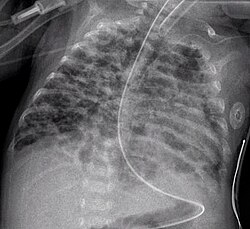
Chest x-ray
A chest X-ray or chest film an x-ray of the chest used to diagnose diseases of the chest. X-rays are among the most common medical images. Doctors use them to diagnose problems.
Like all radiography methods, chest x-rays use ionizing radiation. The chest x-ray process makes a chest x-ray photo. The photo shows the inside of the chest.
Problems identified
|
Chest x-rays are used to find many diseases inside the chest. A doctor can use an x-ray to examine bones, lungs, heart, and great vessels. Pneumonia and congestive heart failure are commonly diagnosed by chest radiograph. Chest x-rays are used to screen for job-related lung disease, for example in mining where workers breathe dust.[1]
Gallery
Chest X-ray Media
Mediastinal structures on a chest radiograph.
A prominent thymus, which can give the impression of a widened mediastinum.
The inferior skin folds of the supraclavicular fossa may give the impression of a periosteal reaction of the clavicle
Projectionally rendered CT scan, showing the transition of thoracic structures between the anteroposterior and lateral view


