File:Sd4hi-unten-crop.jpg
No higher resolution available.
Sd4hi-unten-crop.jpg (398 × 551 pixels, file size: 59 KB, MIME type: image/jpeg)
| This is a file from the Wikimedia Commons. The description on its description page there is shown below.
|
Summary
| Description |
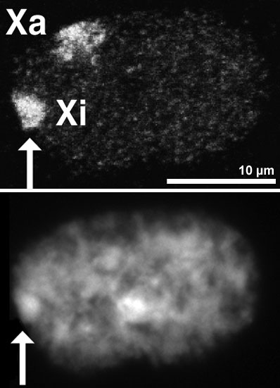
English: Nucleus of a female amniotic fluid cell. Top: Both X-chromosome territories are detected by FISH. Shown is a single optical section made with a confocal microscope. Bottom: Same nucleus stained with Dapi and recorded with a CCD camera. The Barr body is indicated by the arrow, it identifies the inactive X (Xi).
Preparation of specimen as described in: R Eils, S Dietzel, E Bertin, E Schrock, MR Speicher, T Ried, M Robert-Nicoud, C Cremer and T Cremer (1996): Three-dimensional reconstruction of painted human interphase chromosomes: active and inactive X chromosome territories have similar volumes but differ in shape and surface structure. Journal of Cell Biology, Vol 135, 1427-1440. PMID:8978813. Website.
Deutsch: Kern einer weiblichen menschlichen Zelle aus Amnionflüssigkeit. Oben: Darstellung beider X-Chromosomen durch Fluoreszenz-in-situ-Hybridisierung. Gezeigt ist ein einzelner optischer Schnitt, der mit einem konfokalen Laserscanningmikroskop erzeugt wurde. Unten: der gleiche Kern mit Dapi-Färbung, aufgenommen mit einer CCD-Kamera. Das Barr-Körperchen ist hier gut zu erkennen (Pfeil) und identifiziert das inaktive X-Chromosom (Xi).
Präparation wie in: R Eils, S Dietzel, E Bertin, E Schrock, MR Speicher, T Ried, M Robert-Nicoud, C Cremer and T Cremer (1996): Three-dimensional reconstruction of painted human interphase chromosomes: active and inactive X chromosome territories have similar volumes but differ in shape and surface structure. Journal of Cell Biology, Vol 135, 1427-1440. PMID:8978813. Website.
한국어: 여성 양수에 떠있는 세포의 핵. 위: FISH(Fluorescence in situ hybridization)법을 통해 두개의 X염색체를 볼 수 있다. 이 사진은 공초점 레이저 현미경에 의해 찍혔다. 아래 : 같은 핵을 다피 염색(DAPI)법을통해 염색하고, CCD 카메라로 찍은 사진이다. 화살표가 가리키는 것이 바소체이고, Xi로 표시된 것이 불활성화된 X염색체이다(inactive X (Xi).
사진에 관한 내용: R Eils, S Dietzel, E Bertin, E Schrock, MR Speicher, T Ried, M Robert-Nicoud, C Cremer and T Cremer (1996): Three-dimensional reconstruction of painted human interphase chromosomes: active and inactive X chromosome territories have similar volumes but differ in shape and surface structure. Journal of Cell Biology, Vol 135, 1427-1440. PMID:8978813. Website. |
| Date | |
| Source | Steffen Dietzel, Dissertation an der Universität Heidelberg, 1996. (Own work) |
| Author | User:Dietzel65, Steffen Dietzel |
| Permission (Reusing this file) |
I, the copyright holder of this work, hereby publish it under the following license: This file is licensed under the Creative Commons Attribution-Share Alike 3.0 Unported license.
|
File history
Click on a date/time to view the file as it appeared at that time.
| Date/Time | Dimensions | User | Comment | |
|---|---|---|---|---|
| current | 09:53, 1 October 2008 | 398 × 551 (59 KB) | Dietzel65 | {{Information |Description={{en|1=(information to be completed)}} {{de|1=Kern einer weiblichen menschlichen Zelle aus Amnionflüssigkeit. Oben: Darstellung beider X-Chromosomen durch Fluoreszenz-in-situ-Hybridisierung. gezeigt ist ein einzelner optischer |
File usage
The following page uses this file: