File:Spindle centriole - embryonic brain mouse - TEM.jpg

Size of this preview: 481 × 600 pixels. Other resolutions: 192 × 240 pixels | 385 × 480 pixels | 616 × 768 pixels | 821 × 1,024 pixels | 1,283 × 1,600 pixels.
Original file (1,283 × 1,600 pixels, file size: 901 KB, MIME type: image/jpeg)
| This is a file from the Wikimedia Commons. The description on its description page there is shown below.
|
Summary
| Description |
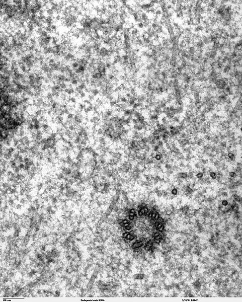
Transmission electron microscope image of a thin section cut through the developing brain tissue (telencephalic hemisphere) of an 11.5 day mouse embryo. This high magnification image of "Embryonic brain 80445" show a spindle centriole and some spindle microtubules visible in the cytoplasm of a mitotic cell at the luminal surface of the telencephalon. JEOL 100CX TEM References: Marin-Padilla, M. (1985) "Early Vascularization of the Embryonic Cerebral Cortex: Golgi and Electron Microscope Studies", J. Comparative Neurology, 241:237-249 Marin-Padilla, M. and M. Amievo (1989) "Early Neurogenesis of the Mouse Olfactory Nerve: Golgi and Electron Microscope Studies", J. Comparative Neurology, 288:339-352 |
| Source | |
| Author | Louisa Howard, Miguel Marin-Padilla |
| Permission (Reusing this file) |
PD |
Licensing
| This work has been released into the public domain by its author, Louisa Howard, Miguel Marin-Padilla. This applies worldwide. In some countries this may not be legally possible; if so: Louisa Howard, Miguel Marin-Padilla grants anyone the right to use this work for any purpose, without any conditions, unless such conditions are required by law.
|
File history
Click on a date/time to view the file as it appeared at that time.
| Date/Time | Dimensions | User | Comment | |
|---|---|---|---|---|
| current | 15:02, 2 November 2006 | 1,283 × 1,600 (901 KB) | Patho | {{Information |Description=Transmission electron microscope image of a thin section cut through the developing brain tissue (telencephalic hemisphere) of an 11.5 day mouse embryo. This high magnification image of "Embryonic brain 80445" show a spindle cen |
File usage
The following page uses this file: