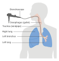
Bronchoscopy
Bronchoscopy is a medical procedure which is used to look at the inside of a person's airways. It can be used to see the inside of the trachea, and the bronchi and bronchioles within the lungs. There are three types of bronchoscopy: rigid, flexible fiberoptic bronchoscopy and CT virtual bronchoscopy (CTVB).
Rigid bronchoscopy
Rigid bronchoscopy is a straight rigid tube used to see into the trachea and proximal bronchi. It is usually done in an operating room under general anesthesia. It is usually used when there is a blockage in either the trachea or the primary bronchi. The diameter of the tube is large enough to insert instruments to remove obstructions.[1]
Flexible fiberoptic bronchoscopy
During the procedure, a thin, flexible fiberoptic tube, called a bronchoscope, is passed through the nose or mouth nose into the airways. The bronchoscope has a light and a camera which lets the doctor see into the airway.[2]
CT virtual bronchoscopy
This technique uses data from multiple CT scans to create a three dimensional (3D) image. This allows doctors to see what is going on without having to operate.[3]
Bronchoscopy Media
References
- ↑ David P. Naidich: Imaging Of The Airways: Functional And Radiologic Correlations. Lippincott Williams & Wilkins; 1 edition (2005) p.30 ISBN 0781757681
- ↑ Pallav Shah: Atlas of Flexible Bronchoscopy. CRC Press; 1 edition (2011) pp. I-II ISBN 034096832X
- ↑ Haliloglu, Mithat; et al. (2003). "CT virtual bronchoscopy in the evaluation of children with suspected foreign body aspiration". European Journal of Radiology. Retrieved 28 March 2013.


