Fallopian tube
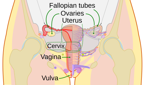
The fallopian tubes (also known as oviducts and uterine tubes) connect the ovaries to the uterus, and let the ovum pass into the uterus where they are able to be fertilized by sperm during sexual intercourse.There are two Fallopian tubes attached to either side of the uterus.[1]
| Fallopian tubes | |
|---|---|
 Schematic frontal view of female anatomy | |
 Cross-section of Fallopian tube, stained and viewed under microscope | |
| Details | |
| System | Reproductive system |
| Artery | tubal branches of ovarian artery, tubal branch of uterine artery via mesosalpinx |
| Lymph | lumbar lymph nodes |
| Identifiers | |
| Latin | Tuba uterina |
| Greek | Salpinx |
| TH | |
| TE | |
| FMA | |
| Anatomical terminology | |
Origin
They are named after the 16th century Italian anatomist, Gabriele Falloppio. The Greek word salpinx (σαλπιγξ) means "trumpet".
Anatomy
There are two Fallopian tubes attached to either side of the end of the uterus. Each tube will end near one ovary. This place is called the fimbria. The Fallopian tubes are not attached to the ovaries, but open into the peritoneal cavity.
In humans, the Fallopian tubes are about 7 - 14 cm long.
Parts
There are four parts of the fallopian tube from the ovary to the uterus:[2]
Layers
The fallopian tube is made of three layers:[3]
- Mucosa - these are folded walls with ciliated cells along them.[4]
- Muscularis externa
- Serosa
Movement
The Fallopian tubes can move around the pelvis.
Fertilization
When an ovum is ready to be released from the ovary, the ovary wall breaks open and the ovum goes into the fallopian tube. There, it starts moving towards to uterus with the help of liquids and cilia on the inside walls. This can take hours or days.
If the ovum is fertilized while in the fallopian tube, then it sticks to the endometrium, which is the beginning of pregnancy.
Related pages
References
- ↑ Williams, J (2018). Williams obstetrics. New York: McGraw-Hill Education Medical. p. 52. ISBN 9781259644320.
- ↑ SUNY Labs 43:04-0101 - "The Female Pelvis: The Oviduct"
- ↑ Fallopian tube
- ↑ Oviduct
