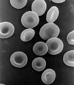
Red blood cell
Red blood cells (also known as RBCs, red blood corpuscles or erythrocytes) are cells in the blood which transport oxygen.[1][2] In women, there are about 4.8 million red blood cells per microliter of blood. In men, there are 5.4 million red blood cells per microliter of blood.[3] Red blood cells are red because they have hemoglobin in them. Quantity of Red Blood Cells in the Human Body. The average male adult has about 5 million red blood cells per cubic millimeter of blood, while the average female adult has about 4.5 million red blood cells per cubic millimeter of blood. This may vary by about 300,000 to 500,000 red blood cells.
Function
The most important function of red blood cells is the transport of oxygen (O2) to the tissues. The hemoglobin absorbs oxygen in the lungs. Then it travels through blood vessels and brings oxygen to all other cells via the heart. The blood cells go through the lungs (to collect oxygen), through the heart (to give all cells oxygen). They go back to the heart to be re-pumped to the lungs (to again collect oxygen), so the blood in your body travels in a double circuit, going through your heart twice before it completes one full circulation of the body.
Red blood cells are doughnut-shaped, but without the hole. This shape is called a bi-concave disc. However, hereditary diseases such as sickle-cell disease can cause them to change shapes and stop blood flow in capillaries and veins. Plasma is got from whole blood. To prevent clotting, an anticoagulant (such as citrate) is added to the blood immediately after it is taken.
Discovery
Jan Swammerdam a scientist from the Netherlands was the first person to observe red blood cells under a microscope. Antoni van Leeuwenhoek, another scientist from the Netherlands, was the first to draw an illustration of "Red Blood Cells".
Discussion
Mammalian RBCs are unique in that they have no cell nucleus in their mature form. These cells have nuclei during development, but push them out as they mature. This gives more space for haemoglobin. Mammalian RBCs also lose all other cellular organelles such as their mitochondria, Golgi apparatus and endoplasmic reticulum. All other vertebrates have nucleated red blood cells.
As a result of not having mitochondria, the cells use none of the oxygen they carry. Instead they produce the energy carrier ATP. Because they lack nuclei and organelles, mature red blood cells do not contain DNA and cannot synthesize any RNA. They cannot divide, and have limited repair capabilities.[4] This also makes sure no virus can target mammalian red blood cells.[5]
Transport of CO2 in the blood
Carbon dioxide (CO2) is carried in blood in three different ways. The exact percentages vary depending whether it is arterial or venous blood.
- Most of it (about 68% to 83%) is converted to bicarbonate ions HCO−
3 by the enzyme carbonic anhydrase in the red blood cells.[6] by the reaction CO2 + H2O → H2CO3 → H+ + HCO−
3. - 5% – 10% is dissolved in the blood plasma.[6]
- 5% – 10% is bound to haemoglobin as carbamino compounds.[6]
Red Blood Cell Media
Typical mammalian red blood cells: (a) seen from surface; (b) in profile, forming rouleaux; (c) rendered spherical by water; (d) rendered crenate (shrunken and spiky) by salt. (c) and (d) do not normally occur in the body. The last two shapes are due to water being transported into, and out of, the cells, by osmosis.
Scanning electron micrograph of blood cells. From left to right: human red blood cell, thrombocyte (platelet), leukocyte.
Two drops of blood are shown with a bright red oxygenated drop on the left and a darker red deoxygenated drop on the right.
Animation of a typical human red blood cell cycle in the circulatory system. This animation occurs at a faster rate (~20 seconds of the average 60-second cycle) and shows the red blood cell deforming as it enters capillaries, as well as the bars changing color as the cell alternates in states of oxygenation along the circulatory system.
- RBC Membrane Proteins SDS-PAGE gel.jpg
Red blood cell membrane proteins separated by SDS-PAGE and silverstained
- RBC membrane major proteins.png
Red blood cell membrane major proteins
Affected by Sickle-cell disease, red blood cells alter shape and threaten to damage internal organs.
References
- ↑ Bradfield, Phil; Potter, Steve (2009). Edexcel IGCSE Biology Student Book. Pearson Education. ISBN 9780435966881.
- ↑ Liang, Barbara. "General Anatomy & Physiology: Red Blood Cells". Wisc-Online. Retrieved 2011-03-22.
- ↑ "What Is a Complete Blood Count?". WebMD.
- ↑ Lua error in Module:Citation/CS1/Identifiers at line 630: attempt to index field 'known_free_doi_registrants_t' (a nil value).
- ↑ Zimmer, Carl (2007-03-27). Scientists explore ways to lure viruses to their death. https://www.nytimes.com/2007/03/27/science/27viral.html. Retrieved 2013-03-26.


