Magnetic resonance imaging
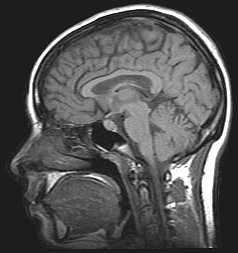
Magnetic resonance imaging (MRI), or nuclear magnetic resonance imaging (NMRI), are techniques that doctors use to give a visual representation of soft tissue (flesh) inside the body. Magnetic resonance uses nuclear magnetic resonance to generate these images.
To take an MRI image, the patient lies on a movable bed. The bed enters a strong magnetic field and then radio waves are applied for a short time in a different direction. This sudden shift causes certain atoms in the patient's body to make special signals. The MRI scanner detects those special signals. The MRI scanner then sends the signal information to a computer, and the computer creates an image of the inner body by using the signal information.
Pros and cons
The MRI is used to diagnose disorders of the body that cannot be seen by X-rays. The MRI is painless and has the advantage of avoiding the dangerous X-ray radiation.
It is an expensive medical procedure, due to the high cost of the equipment. Some metallic objects may distort the image, like hip, shoulder and knee replacements but the exam can still be done. Cochlear (ear) implants, some (older) aneurysm clips in the brain, and most pacemakers are not MRI compatible. Obese and severely claustrophobic patients often need sedation and/or imaging with an "open" magnet, which is much less confining. Contrast agents may or may not be indicated for some patients, and those with compromised kidney function may not be able to receive the contrast, due to a serious but rare skin reaction.
Which parts of the body MRI scans study
MRI scans can be used to study the brain, spinal cord, bones, joints, breasts, the heart and blood vessels.[1] It can also be used to look at other internal organs. MRI scans can be used to find blood clots as well.
An MRI scan can be used as an extremely accurate method of disease detection throughout the body.
Neurosurgeons use an MRI scan not only in defining brain anatomy but in evaluating the integrity of the spinal cord after an injury. An MRI scan can evaluate the structure of the heart and aorta, where it can detect aneurysms or tears. Furthermore, MRI is nowadays used intraoperatively. [2]
It provides valuable information on glands and organs within the abdomen, and accurate information about the structure of the joints, soft tissues, and bones of the body. Often, surgery can be deferred or more accurately directed after knowing the results of an MRI scan.
Magnetic Resonance Imaging Media
- Structural MRI animation.ogv
An animation of MRI images of a human head. For additional information contact Dwayne Reedme on his talk page.
- Mri scanner schematic labelled.svg
Schematic of construction of a cylindrical superconducting MR scanner
- TR TE.jpg
Effects of TR and TE on MR signal
- T1t2PD.jpg
Examples of T1-weighted, T2-weighted and PD-weighted MRI scans
- Siemens Magnetom Aera MRI scanner.jpg
Patient being positioned for MR study of the head and abdomen
- White Matter Connections Obtained with MRI Tractography.png
MRI diffusion tensor imaging of white matter tracts
- PAPVR.gif
MR angiogram in congenital heart disease
Real-time MRI of a human heart at a resolution of 50 ms
Related pages

References
- ↑ "MRI scan - Why it is used". National Health Service. Retrieved September 20, 2012.
- ↑ Lua error in Module:Citation/CS1/Identifiers at line 630: attempt to index field 'known_free_doi_registrants_t' (a nil value).
Further reading
- TRTF/EMRF: The history of MRI (Peter A. Rinck, ed). url = http://www.magnetic-resonance.org/ch/20-01.html
- Guadalupe Portal; Aliosvi Rodriguez Whole body magnetic resonance imaging in early diagnosis in Trinidad BMJ (2010) ISSN 1756-1833 url = http://www.bmj.com/rapid-response/2011/12/19/re-whole-body-magnetic-resonance-imaging
- Lua error in Module:Citation/CS1/Identifiers at line 630: attempt to index field 'known_free_doi_registrants_t' (a nil value).
- Simon, Merrill; Mattson, James S (1996). The pioneers of NMR and magnetic resonance in medicine: The story of MRI. Ramat Gan, Israel: Bar-Ilan University Press. ISBN 0-9619243-1-4.
- Haacke, E Mark; Brown, Robert F; Thompson, Michael; Venkatesan, Ramesh (1999). Magnetic resonance imaging: Physical principles and sequence design. New York: J. Wiley & Sons. ISBN 0-471-35128-8.
- Lua error in Module:Citation/CS1/Identifiers at line 630: attempt to index field 'known_free_doi_registrants_t' (a nil value).
- P Mansfield (1982). NMR Imaging in Biomedicine: Supplement 2 Advances in Magnetic Resonance. Elsevier. ISBN 9780323154062.
- Eiichi Fukushima (1989). NMR in Biomedicine: The Physical Basis. Springer Science & Business Media. ISBN 9780883186091.
- Bernhard Blümich; Winfried Kuhn (1992). Magnetic Resonance Microscopy: Methods and Applications in Materials Science, Agriculture and Biomedicine. Wiley. ISBN 9783527284030.
- Peter Blümer (1998). Peter Blümler, Bernhard Blümich, Robert E. Botto, Eiichi Fukushima (ed.). Spatially Resolved Magnetic Resonance: Methods, Materials, Medicine, Biology, Rheology, Geology, Ecology, Hardware. Wiley-VCH. ISBN 9783527296378.
{{cite book}}: CS1 maint: multiple names: editors list (link) - Zhi-Pei Liang; Paul C. Lauterbur (1999). Principles of Magnetic Resonance Imaging: A Signal Processing Perspective. Wiley. ISBN 9780780347236.
- Franz Schmitt; Michael K. Stehling; Robert Turner (1998). Echo-Planar Imaging: Theory, Technique and Application. Springer Berlin Heidelberg. ISBN 9783540631941.
- Vadim Kuperman (2000). Magnetic Resonance Imaging: Physical Principles and Applications. Academic Press. ISBN 9780080535708.
- Bernhard Blümich (2000). NMR Imaging of Materials. Clarendon Press. ISBN 9780198506836.
- Jianming Jin (1998). Electromagnetic Analysis and Design in Magnetic Resonance Imaging. CRC Press. ISBN 9780849396939.
- Imad Akil Farhat; P. S. Belton; Graham Alan Webb; Royal Society of Chemistry (Great Britain) (2007). Magnetic Resonance in Food Science: From Molecules to Man. Royal Society of Chemistry. ISBN 9780854043408.
Other websites
- A Guided Tour of MRI: An introduction for laypeople Archived 2013-04-12 at the Wayback Machine National High Magnetic Field Laboratory
- Video: What to Expect During Your MRI Exam from the Institute for Magnetic Resonance Safety, Education, and Research (IMRSER)
- 3D Animation Movie about MRI Exam Archived 2009-02-26 at the Wayback Machine
- Magnetic resonance imaging -Citizendium


