Mycobacterium
Mycobacterium is a genus of bacteria, with about 190 species.The genus includes pathogens known to cause serious diseases in mammals, including tuberculosis (Mycobacterium tuberculosis) and leprosy (Mycobacterium leprae).[1] Mycobacteria are immobile and are rod-shaped. They are mostly found in water, soil, and dust.
| Mycobacterium | |
|---|---|

| |
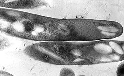
| Transmission electron microscopy (TEM) micrograph of Mycobacterium tuberculosis. | |
| Scientific classification | |
| Kingdom: | |
| Phylum: | |
| Order: | |
| Suborder: | Corynebacterineae
|
| Family: | Mycobacteriaceae
|
| Genus: | Mycobacterium
|
The colony colour ranges from white to orange to pink (Iivanainen, 1999[2]). Most mycobacteria need oxygen to survive (aerobic organisms), although some species require very little amounts of oxygen, less than what is present in the air, are called (microaerophilic bacteria) (Falkinham, 1996[3]).
Categorization
Mycobacteria can be categorized into three groups:
1.Mycobacterium tuberculosis complex – causative pathogen of tuberculosis
2.Non-tuberculosis mycobacteria (NTM)
3.Mycobacterium leprae – causative pathogen of leprosy
Characterstics
The Greek prefix myco means "fungus". When they are cultured on the surface of liquids, mycobacteria growth is similar to that of moulds, a kind of fungus.[4]
Mycobacteria have an external membrane, contain no layer that lies outside the bacteria (capsules). The special characteristic about all Mycobacterium species is having a thicker cell wall than many other bacteria, which is to maintain the shape of the plasma membrane. The cell wall is waxy and rich in specific types of fats (lipids), called mycolic acids or mycolates.
Mycobacterium tuberculosis can occupy different environments. However, they grow so slowly that their duplication time exceeds 18 hours. Surprisingly, mycobacterium leprae have the longest doubling time of all bacteria. In comparison, bacteria E. coli only takes as short as 20 minutes to double.
Clinical Manifestation
Mycobacteria can colonize their hosts without the hosts showing any adverse signs. Many people around the world have infections of M. tuberculosis without showing any signs of it.
Mycobacterial infections are difficult to treat. The organisms are tough due to their cell wall.[5] In addition, they are naturally resistant to a number of antibiotics that disrupt cell-wall building, such as penicillin. With their unique cell wall, they can survive long exposure to acids, alkalis, detergents, oxidative bursts, lysis by complement, and many antibiotics.
Mycobacterial infections are complex diseases and are more common in individuals with weakened immune systems, for example people undergoing treatment for cancer or that are HIV positive. One third of the planet’s population currently carries the M. tuberculosis bacteria. However, only 25 per cent will develop disease. They can cause tuberculosis, nontuberculous mycobacteria (NTM) pulmonary infections, other localized NTM or disseminated infections, leprosy, and chronic ulcers (Buruli ulcer).
Most mycobacteria are susceptible to the antibiotics clarithromycin and rifamycin, but antibiotic-resistant strains have emerged.
Mycobacterium Media
- Model of the Mycobacterial Cell Envelope.png
Model of the Mycobacterium spp. cell envelope with 3-D protein structures
- Three Pathogenic Mycobacteria.png
Orthologous proteins found in each species (based on OMA identifiers). Unique proteins for each species are localized in the outer section for each species.
Phylogenetic tree of slowly-growing members of the Mycobacterium genus
- Phylogentic Tree of Rapidly-Growing Mycobacterium Tortoli 2017.png
Phylogenetic tree of rapidly-growing members of the Mycobacterium genus, alongside the M. terrae complex.
- Mycobacterium tuberculosis Ziehl-Neelsen stain 02.jpg
Mycobacterium tuberculosis on Ziehl-Neelsen stain
- Slant tubes of Löwenstein-Jensen medium with control, M tuberculosis, M avium and M gordonae.jpg
Slant tubes of Löwenstein-Jensen medium.
- Mycobacteria Growth Indicator Tube (MGIT) samples in ultraviolet light.jpg
MGIT samples emitting fluorescence in ultraviolet light
References
- ↑ Sherris medical microbiology : an introduction to infectious diseases (4th ed.). New York: McGraw-Hill. 2004. ISBN 0-8385-8529-9. OCLC 52358530.
- ↑ Lua error in Module:Citation/CS1/Identifiers at line 630: attempt to index field 'known_free_doi_registrants_t' (a nil value).
- ↑ Lua error in Module:Citation/CS1/Identifiers at line 630: attempt to index field 'known_free_doi_registrants_t' (a nil value).
- ↑ James H. Kerr and Terry L. Barrett, "Atypical mycobacterial diseases", Military Dermatology Textbook, p. 401.
- ↑ which is neither truly Gram negative nor positive
6. Antonini, J. (2014) "Sarcoidosis", Reference Module in Biomedical Sciences. Available at: <https://www.sciencedirect.com/topics/medicine-and-dentistry/mycobacterium>
7. Rice, L. and Jung, M. (2018) "Neutrophilic Leukocytosis, Neutropenia, Monocytosis, and Monocytopenia", Hematology, pp. 675–681. Available at: <https://www.sciencedirect.com/topics/medicine-and-dentistry/mycobacterial-infection>