Chemical synapse
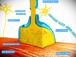
Chemical synapses are synapses that use chemical messengers called neurotransmitters to transmit signals. They are found all over the body. Especially in the central nervous system and the brain.
Neurons use electrical signals to carry information.[1] These signals are called action potentials. There are an estimated 86 billion neurons in the average human brain.[2][3] Neurons don't act alone. They need to connect to other neurons and pass messages between each other. The electrical signal cannot pass the gap between neurons alone. This is why neurotransmitters are needed to pass signals from one neuron to the next. In this sense they differ from electrical synapses which pass electric signals directly to the next neuron.[4] Chemical synapses can be further classified depending upon function and structure.[5]
Structure
The structure of a typical chemical synapse comes in three parts:
- The pre-synaptic terminal is usually on the axon. This releases neurotransmitters into the synaptic cleft. The pre-synaptic terminal is the first part of synaptic transmission and so has a "pre-" prefix.
- The synaptic membrane of the post-synaptic cell is usually on the dendrite of the next neuron. This absorbs neurotransmitters into the post-synaptic neuron (the neuron that is receiving the signal). The post-synaptic cell is the last part of the transmission process and so has a "post-" suffix.
- The synaptic cleft is the bit in the middle of the two membranes. This space is filled with an extracellular (extra- = outside of. cellular = live cell.) matrix of proteins that mainly acts to hold the two neurons together.
Two types of chemical synapse
- Type I synapses are the more common chemical synapse in the human brain.[6] These synapses excite (trigger) the next neuron.[7] The next neuron produces an action potential when this synaptic transmission happens. These synapses are usually found on dendrites of the post-synaptic cell. The pre-synaptic terminal is found on the axon of the neuron that sends the transmission (pre-synaptic cell).[8] Type I synapses are symmetrical in shape.
- Type II synapses are less common in the human brain.[6] These synapses are asymmetrical in shape. They inhibit. Instead of causing an action potential in the next neuron, these synapses stop an action potential. They are less common than type I synapses.[7]
References
- ↑ Neuroscience for kids; Washington University
- ↑ Isotropic Fractionator: A simple, rapid method for the quantification of total cell count; The journal of Neuroscience.
- ↑ Synapse-A primer; "How many synapses in the human brain?" Archived 2014-06-14 at the Wayback Machine; The DANA Foundation
- ↑ Electrical synapses; NCBI books
- ↑ University of Texas, Austin. Synapse web page.
- ↑ 6.0 6.1 Synapses Archived 2014-06-14 at the Wayback Machine; Boston University
- ↑ 7.0 7.1 Chemical synapses Archived 2016-11-30 at the Wayback Machine; Encyclopaedia of Life Science; page 5; John Wiley and Sons.
- ↑ The Synapse-A primer
Chemical Synapse Media
synapsis video clip synopsis
Archived 2014-06-14 at the Wayback Machine; DANA Foundation


