Aphelion (software)
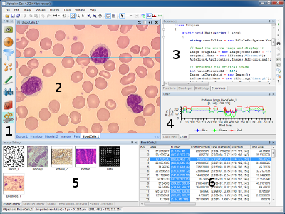
The Aphelion Imaging Software Suite is a set of computer programs for image processing and image analysis. It includes three base products: Aphelion Lab, Aphelion Dev, and Aphelion SDK.
| AphelionDevScreenshot.PNG Aphelion Dev graphical user interface | |
| Developer(s) | ADCIS French company |
|---|---|
| Initial release | 1996 |
| Stable release | 4.6.0 / 27 October 2022 |
| Development status | Active |
| Written in | C sharp, C++ |
| Operating system | Windows |
| Type | Image processing |
| License | Proprietary commercial software |
| Website | www |
The Aphelion software product can be used to prototype applications using the graphical user interface. It provides a set of tools for programmers which have to develop software with image analysis needs.
+{{{1}}}−{{{2}}}
Applications
The Aphelion Imaging Software Suite is used by students, researchers, engineers, and software developers in application domains requiring image processing and computer vision,[1][2] such as:
- security (surveillance, object tracking)
- remote sensing
- quality control for the industry and inspection applications
- materials science
- life sciences (medicine and biology)
- earth science (geology)
- theory (image processing, machine learning and optimization)
Specifications
All products of the Aphelion Imaging Software Suite can be run on PC equipped with Windows (Vista, 7, 8, 8.1,[3] or 10) 32 or 64 bits.[4] An online help[5] and video tutorials are available to the user.[6]
Software extensions
Below is a list of Aphelion optional extensions:[7][8]
- 3D Image Processing and 3D Image Display: A set of extensions to display and process 3D images. The 3D display extension is based on the VTK software product.[9]
- 3D Skeletonization: Extension to compute the 3D skeleton.
- Image Registration: Image registration extension to register images coming from different acquisition devices.
- Classification Tools: Classification extension including a « Fuzzy Logic » (fuzzy logic classification),« Neural Networks » (classification based on neural networks), and « Random Forest » (classification based on random forests, derived from the R software product)
- Kriging: Specific extension to remove image noise using geostatistics techniques.
- Camera interface drivers and microscope interface software
- Virtual Image Capture and Virtual Image Stitcher: Two software products to capture mult-field images and stitch them into one single and very large image in the fields of optical and electron microscopy (image stitching).
- Stereology Analyzer: Software to analyze a very large image using stereology. This extension is mainly used in the field of biology on images acquired by a scan microscope.
- VisionTutor: Online image processing course including all the theory and application macro commands that are compatible with Aphelion.
The Aphelion user can add his/her own macro-commands in the user interface[7] that have been automatically recorded to process a batch of images. He/she can also add plugins and libraries in the GUI that have been developed outside the Aphelion environment.[10]
Software versions
Notes and references
- ↑ "Aphelion application fields". adcis.net. Retrieved 5 November 2015.
- ↑ "Aphelion application fields videos". adcis.net. Retrieved 5 November 2015.
- ↑ "What is new in Aphelion 4.3 release". adcis.net. Retrieved 18 November 2015.
- ↑ "Aphelion Dev overview". adcis.net. Retrieved 18 November 2015.
- ↑ "Help documents". adcis.net. Retrieved 5 November 2015.
- ↑ "ADCIS (Advanced Concepts in Imaging Software) products videos". adcis.net. Retrieved 5 November 2015.
- ↑ 7.0 7.1 "Official Aphelion Dev brochure (2015)" (PDF). adcis.net. Retrieved 19 November 2015.
- ↑ "Software extensions brochures". adcis.net. Retrieved 18 November 2015.
- ↑ Legland, David (March 2009). "Solutions logicielles pour le traitement d'images" [Softwares for addressing image processing] (PDF). INRA (Institut National de la Recherche Agronomique) (in français). Retrieved 25 February 2016.
- ↑ "Aphelion user guide (Loading a macro as an Aphelion plugin)" (PDF). adcis.net. Retrieved 5 November 2015.
- ↑ "Aphelion version and changes history". adcis.net. Retrieved 5 November 2015.
Other references
Miscellaneous
- "Image analysis software list". peipa.essex.ac.uk. Retrieved 19 November 2015.
- "Aphelion 2006 advertising" (PDF). AdvancedImagingPro.com. Archived from the original (PDF) on 13 September 2018. Retrieved 19 November 2015.
Materials science applications
- Hénault, Éric (2006). "Method of Automatic Characterization of Inclusion Population by a SEM-FEG/EDS/Image Analysis System". Jeol News. 41 (73): 22–24. Retrieved 14 November 2015.
A coupling was carried out between a field-emission scanning electron microscope (JEOL, JSM-6500F), an energy-dispersive spectrometer (EDS) (PGT, detector SDD SAHARA) and image analysis software (APHELION).
- Lua error in Module:Citation/CS1/Identifiers at line 630: attempt to index field 'known_free_doi_registrants_t' (a nil value).
- Lua error in Module:Citation/CS1/Identifiers at line 630: attempt to index field 'known_free_doi_registrants_t' (a nil value).
- Moreaud, Maxime (10 April 2007). "Nanotomographie" [Nanotomography] (PDF). cmm.mines-paristech.fr (in français). Archived from the original (PDF) on 3 March 2016. Retrieved 22 February 2016.
[...] une solution complète d'alignement des projections et de reconstruction tomographique 3D a été développée et intégrée à la plateforme Aphelion.
[ [...] a complete projection alignment and 3D reconstruction plug-in was developed and integrated to Aphelion.] - Lua error in Module:Citation/CS1/Identifiers at line 630: attempt to index field 'known_free_doi_registrants_t' (a nil value).
However, they can be defined after simple morphological transformations on the digitized images of the microstructure: a closing is made on the fiber phase of the yarn by using the Aphelion software.
- Lua error in Module:Citation/CS1/Identifiers at line 630: attempt to index field 'known_free_doi_registrants_t' (a nil value).
- Lua error in Module:Citation/CS1/Identifiers at line 630: attempt to index field 'known_free_doi_registrants_t' (a nil value).
- Lua error in Module:Citation/CS1/Identifiers at line 630: attempt to index field 'known_free_doi_registrants_t' (a nil value).
- Lua error in Module:Citation/CS1/Identifiers at line 630: attempt to index field 'known_free_doi_registrants_t' (a nil value).
Specific programs were developed using Aphelion 3.2f (Adcis S.A.) and Matlab software, with image analysis toolbox version 6.0 from Mathworks (Natick, MA).
- Lua error in Module:Citation/CS1/Identifiers at line 630: attempt to index field 'known_free_doi_registrants_t' (a nil value).
Life sciences applications
- (June 2006) "Effect of GSM-900 RFR on HSP expression in brain immune cells" in 28th Annual Meeting of the BEMS. . hal-00161897, version 1.
The level of heat shock protein expression was estimated using image analysis (Aphelion software).
- Lua error in Module:Citation/CS1/Identifiers at line 630: attempt to index field 'known_free_doi_registrants_t' (a nil value).
The ratio between the surface of bisbenzimide staining and the surface of specific immunostaining was measured by using a software Aphelion 3.2 from Adcis (Herouville Saint Clair, France).
- Lua error in Module:Citation/CS1/Identifiers at line 630: attempt to index field 'known_free_doi_registrants_t' (a nil value).
The area resorbed was quantified by Image Analysis using custom software developed using Aphelion ActiveX objects (ADCIS).
- (4–8 July 2005) "Avantages d'une quantification à basse résolution de la vascularisation intratumorale" in 9th conference of the French Microscopy Society (French: [Société Française des Microscopies] Error: {{Lang}}: text has italic markup (help)) (SFµ). .
Une routine de traitement d'images, développée dans l'environnement du logiciel boîte à outils Aphelion (ADCIS) permet de donner une appréciation objective du degré de vascularisation moyen et maximal de la tumeur.
[An image analysis macro-command, developed in the Aphelion (ADCIS) toolbox software, allows to appreciate objectively the mean and maximum grades of tumor vascularisation.] - Le Maire, Sophie; et al. (2005). "Caractérisation par analyse d'images de l'angiogenèse sur des coupes histologiques" [Image analysis quantization of angiogenesis on images of histological slides]. Group for Study of Signal and Image Analysis (French: [GRETSI (Groupe d'Études du Traitement du Signal et des Images)] Error: {{Lang}}: text has italic markup (help)) (in français). hdl:2042/14096. Retrieved 22 February 2016.
{{cite journal}}: Italic or bold markup not allowed in:|journal=(help)Les logiciels utilisés pour réaliser ce travail sont les suivants : (a) APHELION v.3.2 pour le traitement d'images 2D, la reconstruction 3D et les mesures 2D et 3D, [...]
[Softwares used to accomplish this work are: (a) APHELION v.3.2 for 2D image analysis, 3D reconstruction, and 2D and 3D measurements, [...] ] - Ghazi, Kamelia; et al. (16 November 2012). "Hyaluronan Fragments Improve Wound Healing on In Vitro Cutaneous Model through P2X7 Purinoreceptor Basal Activation: Role of Molecular Weight". PLOS ONE. 7 (11): e48351. Bibcode:2012PLoSO...748351G. doi:10.1371/journal.pone.0048351. PMC 3500239. PMID 23173033.
Percentage of wound area was measured using Aphelion Dev image processing and analysis software developed by ADCIS S.A.
- Lua error in Module:Citation/CS1/Identifiers at line 630: attempt to index field 'known_free_doi_registrants_t' (a nil value).
- Lua error in Module:Citation/CS1/Identifiers at line 630: attempt to index field 'known_free_doi_registrants_t' (a nil value).
For image analysis the Aphelion software package was used made by Adcis SA and AAI Inc.
Earth science applications
- Lua error in Module:Citation/CS1/Identifiers at line 630: attempt to index field 'known_free_doi_registrants_t' (a nil value).
- Lua error in Module:Citation/CS1/Identifiers at line 630: attempt to index field 'known_free_doi_registrants_t' (a nil value).
- Gauchat, K.; et al. (2006). "Cristal Size Distribution (CSD) of garnets as function of metallographic grade and composition in black marls of the Nufenen zone" (PDF). Geophysical Research Abstracts. 8.
The 2D garnet distributions and garnet shapes were determined using the Aphelion image analysis program
- (6 February 2003) "Taille et Forme des cristaux de quartz dans une géode du forage EPS1 de Soultz-sous-Forêts" in 25th conference of the French Stereology Society. .
- (13–15 October 2003) "Coupled THM modelling of the stimulated permeable fractures in the near well at the Soultz-sous-Forêts site (France)" in Geoproc 2003, International Conference on Coupled T-H-M-C Processes in Geosystems. 2 : 665–670. DOI:10.1016/S1571-9960(04)80116-2.
Theory applications
- Hanbury, Allan; Serra, Jean (2002). "A 3D-polar coordinate colour representation suitable for image analysis" (PDF). University of Technology, Vienna, Austria. PRIP-TR-77. Retrieved 5 November 2015.
{{cite journal}}: Cite journal requires|journal=(help)Software already used by the author which implement cylindrically shaped colour models include: Matlab release 12.1, Aphelion 3.0, Optimas 6.1 and Paint Shop Pro 7.
- Brambor, Jaromír (11 July 2006) (in fr). Algorithmes de la morphologie mathématique pour les architectures orientées flux. p. 80. http://cmm.ensmp.fr/~brambor/public/docs/Brambor-Doctoral-Thesis/Brambor-Doctoral-Thesis.pdf. Retrieved 23 February 2016.

