Heart
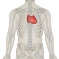
The heart is an organ found in every vertebrate. It is a very strong muscle. It is on the left side of the body in humans and is about the size of a fist. It pumps blood throughout the body. It has regular contractions, or when the heart squeezes the blood out into other parts of the body.
Cardiac and cardio both mean "about the heart", so if something has the prefix cardio or cardiac, it has something to do with the heart.
Myocardium means the heart muscle: 'myo' is from the Greek word for muscle - 'mys', cardium is from the Greek word for heart - 'kardia'.
Structure
The human heart has four chambers or closed spaces. Some animals have only two or three chambers.
In humans, the four chambers are two atria and two ventricles. Atria is talking about two chambers; atrium is talking about one chamber. There is a right atrium and right ventricle. These get blood that comes to the heart. They pump this blood to the lungs. In the lungs blood picks up oxygen and drops carbon dioxide. Blood from the lungs goes to the left atrium and ventricle. The left atrium and ventricle send the blood out to the body. The left ventricle works six times harder than the right ventricle because it carries oxygenated blood.[1]
Blood is carried in blood vessels. These are arteries and veins. Blood going to the heart is carried in veins. Blood going away from the heart is carried in arteries. The main artery going out of the right ventricle is the pulmonary artery. (Pulmonary means about lungs.) The main artery going out of the left ventricle is the aorta.
The veins going into the right atrium are the superior vena cava and inferior vena cava. These bring blood from the body to the right heart. The veins going into the left atrium are the pulmonary veins. These bring blood from the lungs to the left heart.
When the blood goes from the atria to the ventricles it goes through heart valves. When blood goes out of the ventricles it goes through valves. The valves make sure that blood only goes one way in or out.
The four valves of the heart are:
- Atria to ventricle valves
- Tricuspid valve – blood goes from right atrium to right ventricle
- Mitral valve – blood goes from left atrium to left ventricle
- Ventricles to arteries
- Pulmonic valve – blood goes out of the right ventricle to the lungs (through the pulmonic artery)
- Aortic valve – blood goes out of the left ventricle to the body (through the aorta)
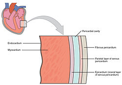
The heart has three layers. The outer covering is the pericardium. This is a tough sack that surrounds the heart. The middle layer is the myocardium. This is the heart muscle. The inner layer is the endocardium. This is the thin smooth lining of the chambers of the heart.[2]
Cardiac cycle
A heart beat is when the heart muscle contracts. This means the heart pushes in and this makes the chambers smaller. This pushes blood out of the heart and into the blood vessels. After the heart contracts and pushes in, the muscle relaxes or stops pushing in. The chambers get bigger and blood coming back to the heart fills them.
When the heart muscle contracts (pushes in) it is called systole. When the heart muscle relaxes (stops pushing in), this is called diastole. Both atria do systole together. Both ventricles do systole together. But the atria do systole before the ventricles. Even though the atrial systole comes before ventricular systole, all four chambers do diastole at the same time. This is called cardiac diastole
The order is: atrial systole → ventricular systole → cardiac diastole. When this happens one time, it is called a cardiac cycle.
Heart's pacemaker
Systole (when the heart squeezes) happens because the muscle cells of the heart gets smaller in size. When they get smaller we also say they contract. Electricity going through the heart makes the cells contract. The electricity starts in the sino-atrial node (acronym SA Node) The SA Node is a group of cells called pacemaker cells in the right atria. These cells start an electrical impulse. This electrical impulse sets the rate and timing at which all cardiac muscle cells contract. This motion is called 'atrial systole'. Once electrical impulse goes through the atrio-ventricular node (AV Node). The AV Node makes the impulse slow down. Slowing down makes the electrical impulses get to the ventricles later. That is what makes the ventricular systole occur after atrial systole, and lets all the blood leave the atria before ventricle contracts (meaning squeeze).
After the electrical impulse goes through the AV Node, the electrical impulse will go through the conduction system of the ventricle. Conduction means heat or electricity traveling through something. This brings the electrical impulse to the ventricles. The first part of the conduction system is the bundle of His. His is named for the doctor (Wilhelm His, Jr) who discovered it. Bundle means strings or wires grouped together in parallel. Once the bundle (meaning a group of strings or wires going in parallel directions) goes through the ventricle muscle, it divides into two bundle branches, the left bundle branch and the right bundle branch. The left bundle branch travels to the left ventricle and the right bundle branch travels to the right ventricle. At the end of the bundle branches, the electrical impulse goes into the ventricular muscle through the Purkinje Fibers. This is what makes ventricle contraction take place and makes ventricular systole.
The order is: Sino-Atrial Node → Atria (systole) → Atrio-Ventricular Node → Bundle of His → Bundle branches → Purkinje Fibers → Ventricles (systole)
ECG
ECG is an acronym for ElectroCardioGram. It is also written EKG for ElectroKardioGram in German. The ECG shows what the electricity in the heart is doing. An ECG is done by putting electrodes on a person's skin. The electrodes see the electricity going through the heart. This is written on paper by a machine. This writing on the paper is the ECG.
Doctors learn about the person's heart by looking at the ECG. The ECG shows some diseases of the heart like heart attacks or problems with the rhythm of the heart (how the electricity goes through the heart's conduction system.)
The ECG shows atrial systole. This is called a P-wave. Then ventricular systole happens. This is called the QRS or QRS-complex. It is called a complex because there are three different waves in it. The Q-wave, R-wave, and S-wave. Then the ECG shows ventricular diastole. This is called the T-wave. Atrial diastole happens then too. But it is not seen separate from ventricular diastole.
The PR-Interval is the space between atrial systole (P) and ventricular systole (QRS). The QT-Interval is from when the QRS starts to when the T ends. The ST-segment is the space between the QRS and T.
Heart Media
Human heart during an autopsy
Cardiology Video
Real-time MRI of the human heart
The human heart is in the middle of the thorax, with its apex pointing to the left.
Frontal section showing papillary muscles attached to the tricuspid valve on the right and to the mitral valve on the left via chordae tendineae.
Blood flow through the heart
Video explanation of blood flow through the heart
References
- ↑ Phibbs, Brendan (2007). The human heart: a basic guide to heart disease (2nd ed.). Philadelphia: Lippincott Williams & Wilkins. p. 1. ISBN 9780781767774.
- ↑ Betts, J. Gordon (2013). Anatomy & physiology. ISBN 978-1938168130. Retrieved 11 August 2014.
Other websites
| Wikimedia Commons has media related to Lua error in Module:Commons_link at line 62: attempt to index field 'wikibase' (a nil value).. |
- Symptoms of heart disease
- What is the heart? - NIH
- The gross physiology of the cardiovascular system (2nd Ed., 2012) – Robert M. Anderson, M.D. (CC-BY-NC)
- Atlas of human cardiac anatomy
- " healthy heart " Blog


